
Karolinska researchers distinguish healthy and diseased immune cells with fluorescent nanosensors
This digest was compiled by AI from multiple sources — links to the originals are below.
Researchers at Karolinska Institutet and SciLifeLab have developed a method to analyze the physical condition of immune cells in blood, distinguishing patients with atherosclerosis from healthy individuals. The technique, spectral biophysical cytometry (SBC), uses fluorescent nanosensors and advanced flow cytometry to measure multiple properties of thousands of cells in minutes. The study, published in Nature Nanotechnology, identified altered cell membrane organization and mitochondrial function in T cells from 38 patients.
Spectral Biophysical Cytometry
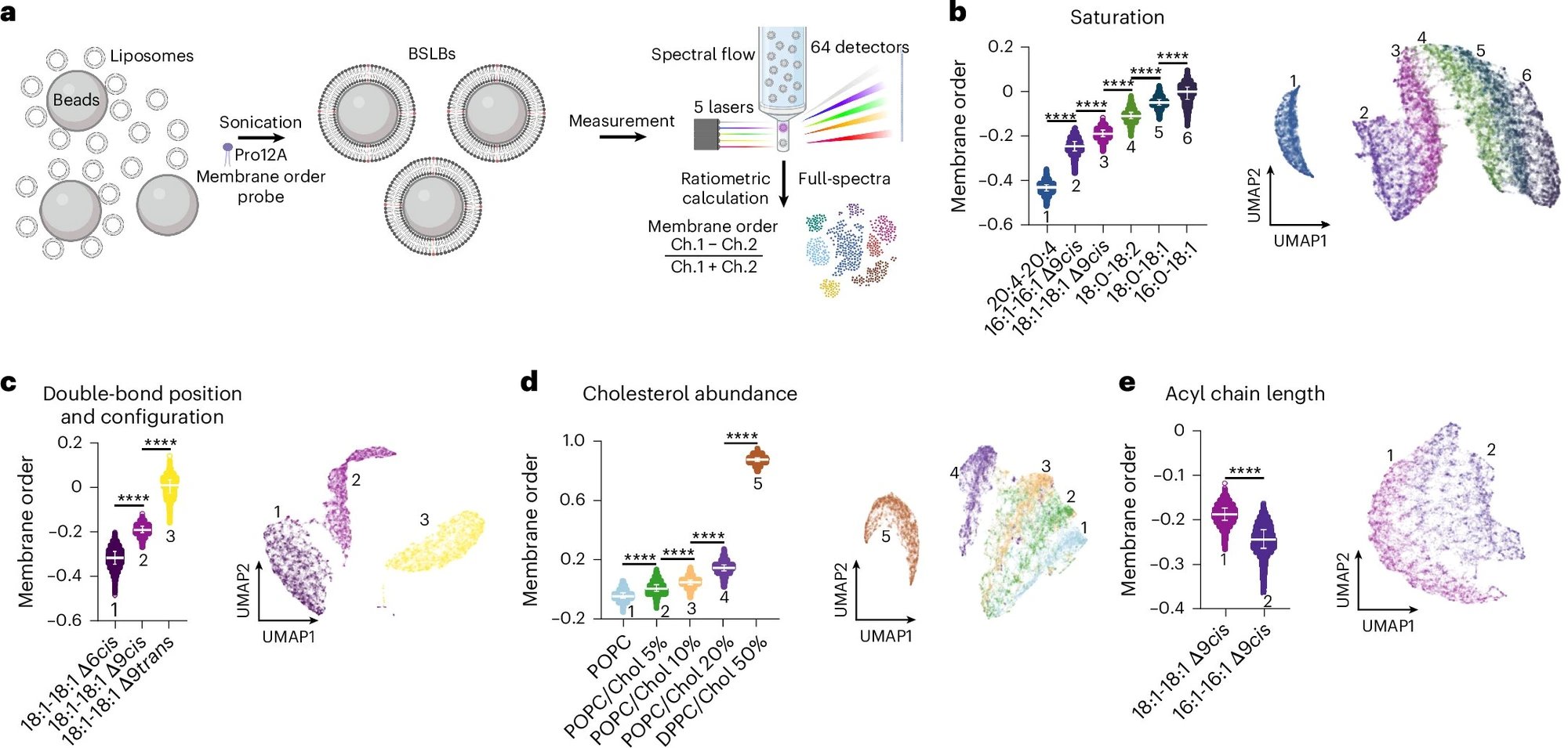
Erdinc Sezgin and colleagues at Karolinska Institutet and SciLifeLab developed SBC to measure physical cell properties like membrane structure and mitochondrial function. The method combines fluorescent nanosensors that react to the cellular environment with high-throughput flow cytometry, analyzing thousands of individual cells in a few minutes. This allows for simultaneous measurement of multiple physical parameters, a capability previously unavailable for large cell populations, and is described in Nature Nanotechnology.
Atherosclerosis Patient Findings
In a study of 38 atherosclerosis patients and 26 controls, SBC revealed significant differences in immune cell physical properties. T cells from patients showed altered membrane organization and mitochondrial function, which correlated with changes in lipid composition and gene activity. These physical changes suggest that cell mechanics play a role in disease processes, and the method can detect them directly in blood samples without lengthy genetic or protein analyses.
Proof-of-Concept Study
The research, led by Sezgin and first author Cenk O. Gurdap, is a proof-of-concept demonstrating SBC's potential. The technique provides an integrated picture of immune cell health, serving as a complement to more time-consuming and costly analyses. It was tested specifically on atherosclerosis but may be applicable to other diseases involving immune cell dysregulation.
1 source
Time · lag behind first