
MIT researchers achieve 0.6-angstrom precision with single-laser microscopy method
This digest was compiled by AI from multiple sources — links to the originals are below.
MIT and Broad Institute researchers have developed a super-resolution imaging technology called U-STORM that achieves localization precision of 0.6 angstroms. The method uses upconverting nanoparticles that blink indefinitely under a single near-infrared laser, eliminating the need for complex imaging buffers and multiple light sources. The achievement, published in Nature Nanotechnology on July 27, overturns the decades-old belief that such nanoparticles are unsuitable for super-resolution microscopy.
The U-STORM Platform
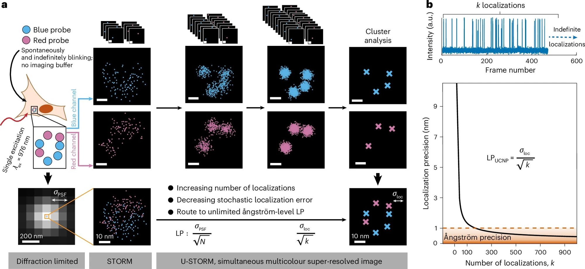
The U-STORM platform relies on compositionally engineered upconverting nanoparticles approximately 10 nm in size that blink spontaneously under continuous near-infrared excitation. Unlike traditional fluorescent dyes, which fade rapidly, these nanoparticles can blink indefinitely, allowing for the collection of over 88,000 localization events from a single particle. The study was led by Sam Peng, the Pfizer Inc.–Gerald Laubach Career Development Assistant Professor of Chemistry at MIT and a core institute member of the Broad Institute. The findings were published in Nature Nanotechnology on July 27.
Precision and Simplicity
U-STORM achieves an unprecedented localization precision of 0.6 angstroms, three orders of magnitude beyond the nanometer-scale limits of standard methods. The technology operates with a single near-infrared laser, which simultaneously excites nanoparticles emitting different colors, unlike conventional multicolor super-resolution imaging that requires multiple expensive lasers and precise optical alignment. This reduces experimental complexity and cost, according to the researchers.
Overturning a Paradigm
For decades, upconverting nanoparticles were considered completely photostable and nonblinking, making them seemingly unsuitable for localization-based super-resolution microscopy. The MIT and Broad Institute team demonstrated that by meticulously controlling nanoparticle composition, they could induce spontaneous blinking without imaging buffers or external optical modulation. This overturns a long-standing assumption and opens new avenues for optical materials in biological imaging.
1 source
Time · lag behind first