
AI use in winning microscopy video sparks scientific-image integrity debate
This digest was compiled by AI from multiple sources — links to the originals are below.
Ning Xu of the National University of Singapore won Nikon's Small World in Motion competition on 15 September with a video of lung cilia. Microscopy experts questioned the biological plausibility of features in the video, suggesting AI may have hallucinated them. Nikon Instruments updated its blog post on 22 September to disclose Xu's use of an unsupervised AI model for post-processing.
Key Facts
- Ning Xu, an optical engineer at the National University of Singapore, won the 2026 Small World in Motion competition on 15 September.
- The winning video shows cilia from a child with primary ciliary dyskinesia, a rare genetic disorder causing chronic lung, sinus and ear infections.
- On 22 September, Nikon Instruments updated its blog post to state that Xu used an unsupervised AI model for post-processing.
- Edward Phelps, a bioengineering researcher at the University of Florida, wrote on LinkedIn that the purple structures resemble mitochondria but such a sub-epithelial structure composed of extracellular mitochondria of the same size as cell nuclei does not occur in biology.
Competition Winner
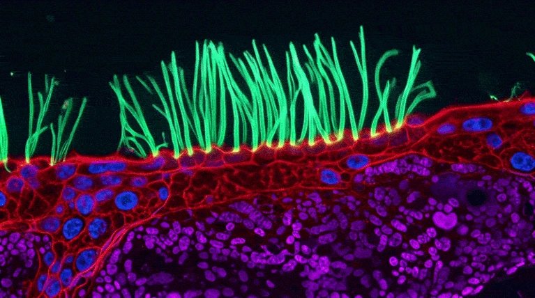
Ning Xu, an optical engineer at the National University of Singapore, was named winner of the annual Small World in Motion competition on 15 September. The competition is run by Nikon Instruments and celebrates videos taken using a light microscope. Xu's entry shows the movement of cilia lining part of the lungs from a child with primary ciliary dyskinesia. In the video, the cilia sit on top of red, purple and blue structures that are not described in the competition's blog post.
Expert Criticism
Days after the announcement, microscopy experts started to question the accuracy of the clip on social media. Edward Phelps, a bioengineering researcher at the University of Florida in Gainesville, wrote on LinkedIn that the purple structures resemble mitochondria but such a sub-epithelial structure composed of extracellular mitochondria of the same size as cell nuclei does not occur in biology. Phelps added that although the blue structures resemble nuclei in appearance, they don't behave like nuclei, and it is unclear what the red stain is supposed to be. Melanie White, a developmental biologist at the University of Queensland, said scientific images are data and researchers need to trust that what they see is grounded in the underlying measurement.
AI Disclosure
On 22 September, Nikon Instruments updated the blog post about Xu's entry to include that he used an unsupervised AI model to assist with post-processing. Xu did not respond to Nature's request for comment about researchers' questions regarding the biological accuracy of his video. Xu wrote on LinkedIn that his team used post-processing to enhance the visual presentation of the regions below the cilia without making anatomical claims about what those rendered features represent. Xu stated that AI was not used to generate the experimental movie, the cilia or their motion.
1 source
Time · lag behind first